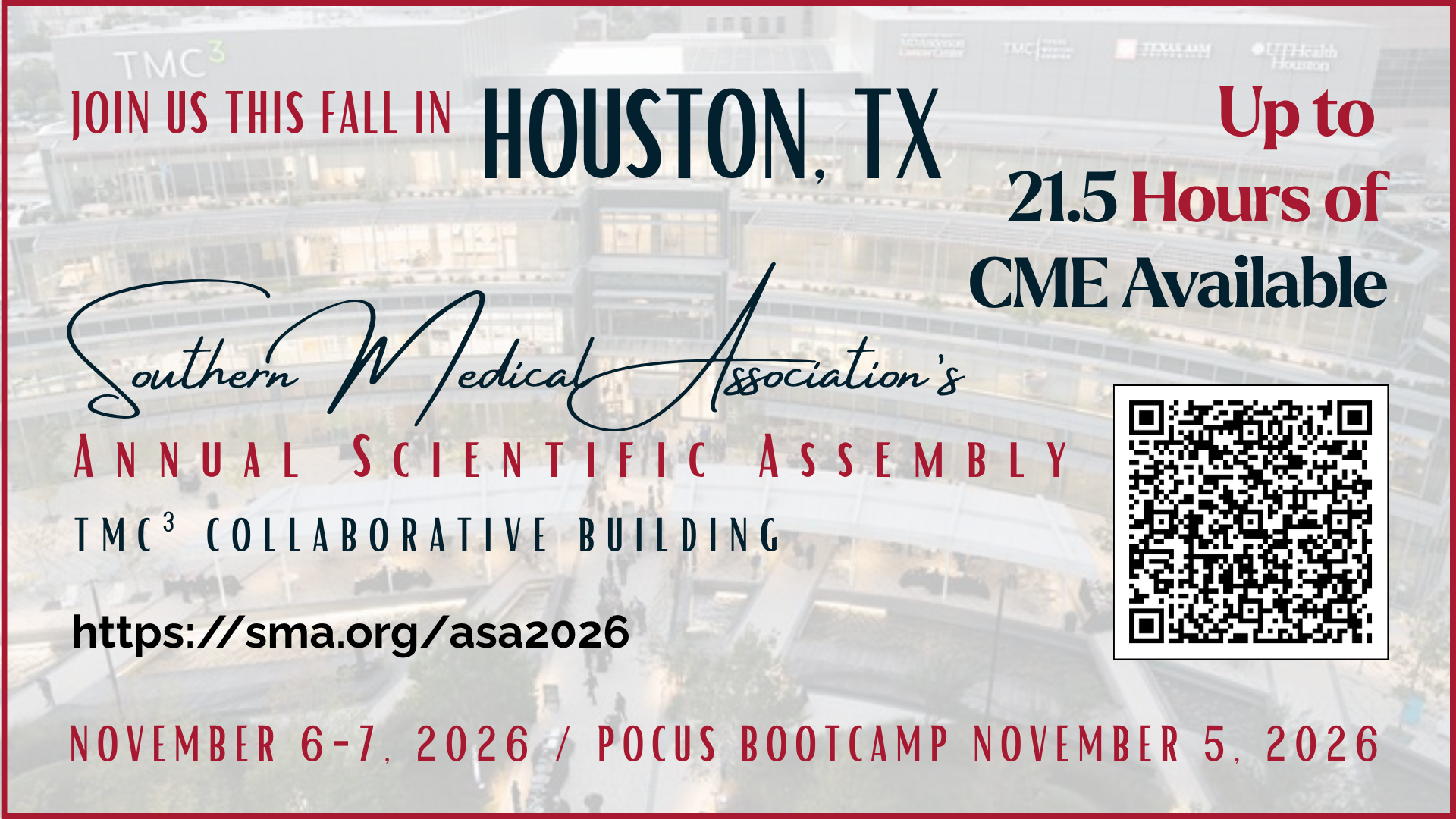
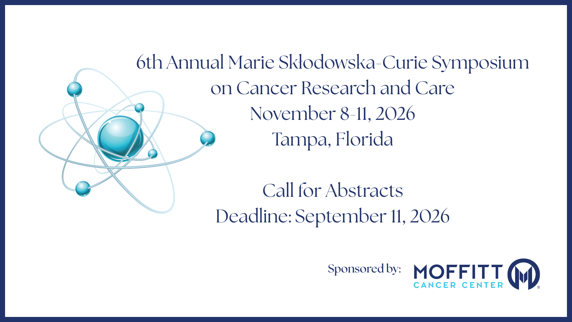
6th Annual Marie Skłodowska-Curie Symposium on Cancer Research and Care: Call for Abstracts

July 16, 2026 // Southern Medical Association

The goal of this symposium is to promote collaboration between cancer care researchers and care providers, biotech, pharma, nonprofit organizations and policymakers from the United States, Canada, and countries of Central and Eastern Europe, to accelerate the development and clinical testing of new cancer treatments and improving cancer care.

Abstract Submission Details

Submit an Abstract

Categories that will be considered:

  • Genomics
  • Immunotherapies
  • Vaccines and Prevention
  • Melanoma
  • Soft Tissue Sarcoma
  • Women’s Health
  • GU
  • GI

Word Limit: 250 words

Submission Format: Oral and Poster - 10-minute presentations each

Submission Instructions: To submit an abstract, you must have an SMA account. If need assistance with creating your free subscriber account, please contact [email protected]. SMA membership is not required to submit an abstract.

Deadline to Submit an Abstract: July 31, 2026, at 11:59 p.m. CDT
Click HERE to submit an abstract.

If you have any questions, please contact:
Caroline Loftis
Southern Medical Association
E-mail: [email protected]
Phone: (205) 421-4177

Posted in: Hidden

Findings: Spotting Acute Cholecystitis on POCUS: What to Look For, What Fools You, and Why it Matters

July 12, 2026 // Caleb Neal, DO, MPH, and Tatiana Havryliuk, MD

Figure 1: Right upper quadrant ultrasound, with a longitudinal view of the gallbladder. Showing a hyperechoic focus with posterior acoustic shadowing (gallstone) in the back of the gallbladder. Anterior gallbladder wall is thickened to 5.1mm, and there is a small rim of pericholecystic fluid in the near field.

POCUS Findings

There are six classic ultrasound findings you're looking for in acute cholecystitis:1,6,8,11,12

  • Gallstones: A bright (hyperechoic) focus inside the gallbladder with a dark shadow behind it, referred to as posterior acoustic shadowing which is the hallmark of a gallstone. Gallstones alone are not indicative of cholecystitis, there must be other signs of infection (included below), so gallstones alone are not included in the six classic findings of acute cholecystitis. 
  • Wall thickening: If the anterior gallbladder wall measures greater than 3mm, then this tells you the gallbladder is inflamed and possibly infected. 
  • Pericholecystic fluid: A thin dark stripe of fluid around the gallbladder, which is indicative of an inflammatory response to a gallbladder infection. 
  • Sonographic Murphy's sign: When the provider pushes the probe directly over the gallbladder, if the patient catches their breath from the tenderness, that's the ultrasound version of Murphy's sign. 
  • Stone-In-Neck (SIN) Sign: A stone impacted in the gallbladder neck or the cystic duct is the root cause of acute cholecystitis, and can be an important indicator if found on ultrasound. 
  • Gallbladder Distention (Hydrops): A normal gallbladder should have a transverse diameter of 4cm or less and a transverse diameter of 10cm or less. Anything greater than 4cm by 10cm is considered abnormal. 
  • Dilated Common Bile Duct (CBD): A normal CBD diameter is less than or equal to 6mm in people under 70, add 1mm to the CBD for every 10 years over 60 (i.e. 70-years-old = 7mm CBD is normal). Dilated CBD may indicate concomitant choledocholithiasis.

Put these together with the clinical picture, pain lasting more than 6 hours, fever, leukocytosis, and you've got acute cholecystitis diagnosed at the bedside.

What a Gallbladder without Cholecystitis Looks Like on POCUS

Figure 2: Ultrasound of a gallbladder in a longitudinal view: thin wall, anechoic (black) bile. Measurement 1 is the anterior gallbladder wall diameter, and measurement 2 is the CBD diameter. Since the gallbladder wall is less than 3mm and the CBD diameter is less than 6mm, both are considered normal. Hyperechoic stones with posterior acoustic shadows are seen.


Figure 3: Ultrasound of a normal gallbladder in a transverse view with a thin GB wall and anechoic bile with no stones visualized. Measurement 1 is the anterior gallbladder wall, which is less than 3mm and therefore within normal limits.

Before you can spot what's wrong, you need to know what's normal. A healthy gallbladder on ultrasound looks like:1,6

  • A thin-walled, pear-shaped structure filled with dark (anechoic) bile
  • The wall should be less than 3mm thick
  • No bright foci or shadows inside the lumen
  • The gallbladder sits just below the liver edge, use the liver as your acoustic window to find it
  • No fluid around the outside of the gallbladder

Why This Matters: Biliary Colic vs. Cholecystitis

Here's the clinical question that POCUS helps answer: Is this just another episode of biliary colic or has this patient crossed the line into acute cholecystitis?

The dividing line is mostly about time and inflammation:

  • Biliary colic occurs when a stone temporarily blocks the cystic duct. The pain peaks within about an hour and usually resolves within 5 hours. The gallbladder isn't inflamed, it's just irritated. On ultrasound, you'll see stones, but the wall looks normal and there's no pericholecystic fluid.4,10
  • Cholecystitis happens when that blockage doesn't clear. The gallbladder stays obstructed, gets inflamed, and starts to swell. Pain lasts more than 6 hours. On ultrasound, you see stones PLUS secondary signs of inflammation: wall thickening, enlarged gallbladder, a stone-in-neck, pericholecystic fluid, a dilated CBD, and a positive sonographic Murphy's sign.1,4,6,10,11,12

This is why history matters so much. A patient who says "I've had this pain for 2 hours that’s coming and going and it's getting better" is a very different story from "this started 10 hours ago and it's only getting worse."

If your patient has had previous episodes of biliary colic, check out the Hello Sono blog on cholelithiasis for a deeper dive into stone detection and biliary colic management: https://www.hellosono.com/hello-sono-blog/pocus-gallstones-ruq-pain-diagnosis

Common Pitfalls

Gallbladder ultrasound is one of the most reliable bedside exams out there, but there are a few traps that catch people. 

Pitfall #1: Wall thickening that isn't cholecystitis

This is the big one. A thick gallbladder wall does NOT automatically mean cholecystitis. Other conditions cause the wall to thicken:9

  • Heart failure: right-sided congestion causes venous engorgement of the gallbladder wall
  • Liver disease/cirrhosis/portal hypertension: same mechanism, venous congestion
  • Acute hepatitis: diffuse inflammation of the liver can thicken the gallbladder wall
  • Hypoalbuminemia (nephrotic syndrome, malnutrition): low protein causes edema everywhere, including the gallbladder wall
  • Ascites: fluid surrounding the gallbladder can make the wall appear thickened
  • Recent eating: a contracted, postprandial gallbladder can make the wall look artificially thick

The key is context. Wall thickening in a patient with right upper quadrant pain, stones, and a positive sonographic Murphy's sign means cholecystitis. Wall thickening in a patient with known cirrhosis and ascites but no focal tenderness is most likely not cholecystitis.

Pitfall #2: Sludge vs. stones

Biliary sludge (thick, echogenic bile) can look like stones at first glance. The difference? Sludge doesn't cast a posterior acoustic shadow, and it layers dependently (meaning it settles to the bottom) without the sharp, bright edges of a true stone. Sludge can be seen in cholecystitis, but it can also show up in patients who are fasting, on TPN, or critically ill, so it's not specific. 

Pitfall #3: Duodenal artifact

Gas in the duodenum, which sits right next to the gallbladder, can create bright echoes that mimic stones. The difference is that duodenal gas moves with peristalsis, doesn't cast a clean shadow, and isn't inside the gallbladder lumen. Scanning in multiple planes can help to sort this out.

Pitfall #4: Missing stones in the gallbladder neck

Stones love to hide in the neck of the gallbladder, the narrowest part that leads to the cystic duct. If you only scan in a long axis, you might miss them. Always rotate to a short axis and fan through the neck carefully.

Pitfall #5: The "WES" sign

When the gallbladder is packed full of stones, you may not see the lumen at all. Instead, you'll see three layers: the gallbladder Wall, the bright Echo of the stones, and the dark Shadow behind them. This is the WES sign (Wall-Echo-Shadow), and it means the gallbladder is full of stones. Don't mistake it for bowel gas.

Gallbladder Wall Changes Can Evolve Over Time

Furthermore, cholecystitis is a dynamic process, and the ultrasound findings evolve over time. A recent study found that among patients who initially had no gallbladder wall changes on their first ultrasound, nearly 20% developed wall thickening on repeat imaging a median of 4 hours later. Patients who developed these changes had longer hospital stays and were more likely to have confirmed cholecystitis.7

If your clinical suspicion is high but the initial POCUS only shows stones without wall thickening, don't completely dismiss cholecystitis. The inflammatory changes may not have had time to develop yet. Consider serial exams, further workup, or immediate outpatient follow-up if the clinical picture doesn't add up.

Evidence: How Good Is POCUS for Cholecystitis?

POCUS isn't perfect for cholecystitis, no single test is, but it is remarkably useful when combined with clinical judgment. A 2024 meta-analysis pooled data from seven studies (1,464 patients) and found that POCUS had a pooled sensitivity of 86% and specificity of 92% for gallbladder disease overall.12 Other studies also found POCUS to be an integral part of the work-up for cholecystitis: 

  • Gallstone detection by POCUS: Sensitivity 90–96%, specificity 88–96%1
  • Overall ultrasound accuracy for cholecystitis: Sensitivity ~81%, specificity ~83%4
  • Gallstones + sonographic Murphy's sign: Positive Predictive Value (PPV) of 92%10
  • Gallstones + wall thickening: PPV of 95%10
  • No stones + negative sonographic Murphy's sign: Negative Predictive Value (NPV) of 95%10
  • CT vs. ultrasound: A 2024 meta-analysis found comparable diagnostic performance between CT and ultrasound for cholecystitis (CT sensitivity 84% vs. US 79%, p = 0.44), with 82% concordance between the two modalities.2

One study offered an important counterpoint. In their single-center study of 147 patients, POCUS had a sensitivity of only 40% for cholecystitis, significantly lower than the pooled estimates.11 Their study highlighted that POCUS accuracy is operator-dependent and that clinical findings and lab data should always be part of the diagnostic equation. 

The Bedside Sonographic Acute Cholecystitis (SAC) Score combines three clinical findings (postprandial symptoms, RUQ tenderness, and Murphy's sign) with two POCUS findings (gallstones and wall thickening) into a single score. A SAC score below 2 has 100% sensitivity (great for ruling out), while a score of 7 or above has 96% specificity (great for ruling in).5

The bottom line: POCUS won't catch every case of cholecystitis, but when you combine what you see on ultrasound with the clinical picture, it's a remarkably effective bedside tool. A recent large multicenter study reinforced that POCUS should not be used as a standalone diagnostic test, it works best when integrated with clinical and laboratory data.8 And there's a practical throughput benefit too: bedside POCUS by emergency physicians decreases length of stay by 7% overall and up to 15% during evening and overnight hours compared to waiting for a radiology ultrasound.1

Case Resolution

Given the combination of persistent right upper quadrant pain, positive Murphy's sign, leukocytosis, and POCUS findings of gallstones with wall thickening and pericholecystic fluid, the provider diagnoses acute cholecystitis at the bedside.

The patient is made NPO, started on IV fluids and antibiotics, and surgery is consulted. She undergoes laparoscopic cholecystectomy the following morning, within 24 hours of diagnosis. Surgical pathology confirms acute cholecystitis. She's discharged home the next day.

Impact of POCUS

In this case, bedside ultrasound:

  • Confirmed the diagnosis in minutes: no waiting for radiology availability
  • Accelerated the surgical consultation: the surgeon was called before the formal ultrasound was even ordered
  • Reduced time to definitive care: early cholecystectomy within the optimal 1–3 day window
  • Avoided unnecessary additional imaging: no CT was needed
  • Improved patient experience: faster answers, faster treatment, and shorter hospital stay

Conclusion

Acute cholecystitis is one of the most common surgical emergencies, affecting approximately 200,000 people in the US each year.4 POCUS puts the diagnosis in your hands. By combining gallstone detection with secondary signs of inflammation (wall thickening, pericholecystic fluid, sonographic Murphy's sign) and correlating with the clinical picture, you can make a confident bedside diagnosis that changes management in real time.

The key is remembering that no single finding rules cholecystitis in or out. It's the combination of what you see, what the patient feels, and what the labs show that gets you to the answer. And when the picture is clear, don't wait, get surgery involved early. The evidence is unambiguous: early cholecystectomy wins.

References

  1. American College of Emergency Physicians (ACEP) Sonoguide. Gallbladder. www.acep.org. https://www.acep.org/sonoguide/basic/gallbladder
  2. de Oliveira GS, Torri GB, Gandolfi FE, et al. Computed tomography versus ultrasound for the diagnosis of acute cholecystitis: a systematic review and meta-analysis. Eur Radiol. 2024;34(11):6967-6979. doi:10.1007/s00330-024-10783-8
  3. Fugazzola P, Podda M, Tian BW, et al. Clinical Update on Acute Cholecystitis and Biliary Pancreatitis: Between Certainties and Grey Areas. EClinicalMedicine. 2024;78. doi:10.1016/j.eclinm.2024.102929
  4. Gallaher JR, Charles A. Acute Cholecystitis: A Review. JAMA. 2022;307(9):916-924. doi:10.1001/jama.2022.2350
  5. Graglia S, Shokoohi H, Loesche MA, et al. Prospective Validation of the Bedside Sonographic Acute Cholecystitis Score in Emergency Department Patients. Am J Emerg Med. 2021;44:36-40. doi:10.1016/j.ajem.2020.12.081
  6. Hello Sono. POCUS Implementation & Credentialing Support. POCUS Implementation & Credentialing Support. Published November 19, 2025. https://www.hellosono.com/hello-sono-blog/pocus-gallstones-ruq-pain-diagnosis
  7. Ivanov D, Cannata D, Chin KA, et al. Evolution of Secondary Findings in Acute Cholecystitis: A Temporal Analysis From Point-of-Care Ultrasound to Subsequent Imaging. J Emerg Med. 2025. doi:10.1016/j.jemermed.2025.03.009
  8. Nipitkul N, Kotnarin R. Agreement between point-of-care ultrasonography and the Tokyo guidelines 2018 for acute cholecystitis (PACED study). CJEM. 2026;28(6):588-594. doi:10.1007/s43678-026-01161-y
  9. Rosselli, M., Radzina, M. and Lim, A.K.P. (2023). Ultrasound of the Biliary System. In Liver Ultrasound (eds A.K.P. Lim and M. Rosselli). https://doi.org/10.1002/9781119612650.ch6
  10. Strasberg SM. Clinical practice. Acute calculous cholecystitis. N Engl J Med. 2008;358(26):2804-2811. doi:10.1056/NEJMcp0800929
  11. Wehrle CJ, Talukder A, Tien L, Parikh S, Devarakonda A, Holsten SB, Fox ED, Lawson A. The Accuracy of Point-of-Care Ultrasound in the Diagnosis of Acute Cholecystitis. Am Surg. 2022 Feb;88(2):267-272. doi: 10.1177/0003134821989057. Epub 2021 Jan 31. PMID: 33517707.
  12. Wu X, Li K, Kou S, Wu X, Zhang Z. The Accuracy of Point-of-Care Ultrasound in the Detection of Gallbladder Disease: A Meta-analysis. Acad Radiol. 2024 Apr;31(4):1336-1343. doi: 10.1016/j.acra.2023.09.029. Epub 2023 Oct 12. PMID: 37838525.


About the Authors
Dr. Caleb Neal is an emergency medicine resident in NYC passionate about POCUS.

Dr. Tatiana Havryliuk is an emergency physician with over 15 years of clinical POCUS experience, former Emergency Ultrasound Director at The Brooklyn Hospital Center (NY), and founder of Hello Sono. She is dedicated to integrating POCUS into primary care to enhance diagnostic accuracy and patient safety.

LinkedIn: https://www.linkedin.com/in/tatiana-havryliuk-md/
Email: [email protected]

*POCUS images and graphics provided courtesy of Hello Sono.

Posted in: Hidden

Spotting Acute Cholecystitis on POCUS: What to Look For, What Fools You, and Why it Matters

July 12, 2026 // Caleb Neal, DO, MPH, and Tatiana Havryliuk, MD

Case Presentation

A 45-year-old woman with no past medical history presents to the emergency department with right upper quadrant pain that started about 10 hours ago, shortly after dinner. She's had intermittent episodes like this before, usually after eating fatty or greasy foods, but they always dissipated within a few hours. This time, the pain hasn't subsided and has been constant since it started. She also endorses nausea and had one episode of non-bilious, non-bloody emesis. She looks very uncomfortable sitting on the stretcher. 

Vitals: HR 98, BP 134/82, temp 38.2°C, O₂ sat 99% on room air. On exam, she's got significant right upper quadrant tenderness with guarding and a positive Murphy's sign. Labs come back with a WBC count of 13,200 and normal liver enzymes. 

What would you expect to see on ultrasound? And what's the diagnosis?

Figure 1: Right upper quadrant ultrasound, with a longitudinal view of the gallbladder.

Solve This Case

Posted in: POCUS

Inside SMA’s 2026 Annual Scientific Assembly: NASA Astronaut Dr. Cady Coleman

July 10, 2026 // Southern Medical Association

Register for the Assembly

In this special preview episode for SMA’s Annual Scientific Assembly in Houston taking place November 6–7, 2026, SMA President Dr. David Netscher is joined by retired NASA astronaut, Air Force Colonel, and scientist Cady Coleman, PhD, for an inspiring conversation about teamwork, leadership, resilience, and the pursuit of excellence in high-stakes environments.

Drawing from her experiences aboard the Space Shuttle and International Space Station, Dr. Coleman reflects on the years of preparation behind every successful mission and the importance of communication, trust, and adaptability when lives depend on a team working together. She shares stories from astronaut training, Antarctic expeditions, and remote surgical simulations, drawing compelling parallels between space exploration and the practice of medicine.

The discussion explores the value of purposeful checklists, learning from mentors, balancing professional and personal responsibilities, and embracing diverse perspectives to solve complex challenges. Dr. Coleman also offers thoughtful insights into maintaining purpose after achieving major career milestones and why the greatest accomplishments—whether in space or health care—are never the work of one individual alone.

This episode offers a preview of what’s ahead at the Assembly, where Dr. Coleman will present "Mental & Physical Lessons Learned from Long Endurance Space Flight," sharing practical lessons on collaboration, leadership, and innovation that physicians can apply to their own careers and patient care.

Learn more and register by clicking on the button below or by calling (800) 423-4992. We hope to see you in Houston!

Register for the Assembly

Posted in: Annual Scientific Assembly

“A Practical Guide to Renal and Bladder Ultrasound” — Southern Medicine Podcast is Now Available

July 9, 2026 // Southern Medical Association

In this episode of the “Southern Medicine Podcast,” Tatiana Havryliuk, MD, presents a focused session on using ultrasound to evaluate suspected renal colic, urinary retention, and other common genitourinary presentations in everyday practice.

Dr. Havryliuk also demonstrates when renal and bladder ultrasound is most useful, how to obtain basic kidney and bladder views, measure bladder volume at the bedside, and recognize normal anatomy. In addition, she reviews key findings such as hydronephrosis and urinary retention, showing how point-of-care ultrasound can guide safe clinical decision-making and help reduce unnecessary imaging.

Dr. Tatiana Havryliuk is an emergency physician and founder of Hello Sono. With 15 years of experience using POCUS in diverse settings, from urban EDs to Everest Base Camp, she is dedicated to extending its benefits to more patients. With Hello Sono, she and her team support practices in building compliant, high-quality, and profitable POCUS programs through tailored education and implementation.

Posted in: POCUS

IMG Spotlight: Arsalan Zafar Iqbal

July 6, 2026 // Southern Medical Association

My IMG Story
By Arsalan Zafar Iqbal, MD

I am an International Medical Graduate from Pakistan, and my journey into medicine has been shaped by curiosity, persistence, and a deep desire to train within a system known for its structured learning and commitment to evidence-based care. I completed my medical education at FMH College of Medicine & Dentistry in Lahore, where I first began to imagine the possibility of building a career in the United States.

From the beginning, the path was neither straightforward nor clearly defined. Like many IMGs, I entered a process where guidance was limited, expectations were often unclear, and much of the journey had to be self-directed. Balancing clinical training, USMLE preparation, and long-term planning required constant adaptation and resilience. Each step forward came with uncertainty, but also with a growing sense of purpose.

My journey to the United States began with my first role as an Emergency Department Technician at the University of Mississippi Medical Center. It was here that I was introduced to the pace, structure, and complexity of the U.S. healthcare system. Working in this environment while still navigating the early stages of my IMG journey taught me the importance of humility, observation, and learning from every clinical interaction, no matter the role.

Soon after, I transitioned into postdoctoral research training at the University of Mississippi, where I began building a stronger academic foundation. This experience opened doors to further opportunities at the University of Michigan, where I worked in a clinical research fellowship in gastroenterology alongside mentors who deeply influenced my growth as a physician and researcher. These experiences also allowed me to contribute to research presented at national meetings, including ACG, DDW, and AASLD, strengthening my commitment to academic medicine and inquiry-driven care.

Being an IMG also means learning to adapt to a new professional culture while being far from the comfort of home, family, and familiarity. Early on, everything felt new—systems, expectations, communication styles, and even the pace of decision-making. Over time, with the guidance of mentors and the support of colleagues, those unfamiliar environments gradually became spaces of growth and belonging.

Throughout this journey, mentorship played a defining role. I was fortunate to learn from individuals who not only guided my academic development but also shaped my perspective on leadership, collaboration, and integrity in medicine. Their support reinforced a core belief that continues to guide me today: success in medicine is rarely achieved alone.

As I reflect on my path, I recognize that the IMG journey is rarely linear. It is built on persistence through uncertainty, resilience in the face of setbacks, and the willingness to continue moving forward even when the path is unclear. Each stage of the journey contributes not only to professional growth, but also to personal transformation.

Today, I am honored to serve as the Lead of the Education Subcommittee for the International Medical Graduate (IMG) Support & Advocacy Ad Hoc Committee at the Southern Medical Association. In this role, I am committed to leading, guiding, supporting, and inspiring IMGs as they navigate their journey toward a career in U.S. medicine. My goal is to help create educational resources, mentorship opportunities, and supportive pathways that empower IMGs to succeed and exceed expectations.

One of the most meaningful lessons I carry with me comes from my mentor, Dr. William D. Chey, who often reminds his trainees that “There are lots of ways to make a difference.” This philosophy continues to guide my approach to medicine, mentorship, and advocacy. Through my work with the Southern Medical Association, I hope to inspire fellow IMGs to recognize their own potential to lead, to serve, and to create meaningful change within medicine.

Dr. Iqbal is an Internal Medicine resident at Magnolia Regional Health Center and serves as the leader of SMA's IMG Support & Advocacy Ad Hoc Committee Education Subcommittee.
Posted in: IMG

100 Words: SMA President Dr. David Netscher’s July Message

July 1, 2026 // David Netscher, MD

I saw a hospital patient with digital ischemia. My hand-held Doppler examination revealed normal arterial flow from forearm to hand. She also reported shortness of breath, and her finger skin appeared thickened. Embracing a multidisciplinary approach, our rheumatologist, internist, pulmonologist, and I diagnosed systemic sclerosis with pulmonary fibrosis and initiated CellCept, sildenafil, and Botox therapy.

It has been my privilege to interview several featured Scientific Assembly speakers, who likewise emphasize multidisciplinary care. Preview Dr. John Elfar's "Interdisciplinary Collaboration Improves Clinical Outcomes" and "Get Out of Your Lane", plus Dr. John Fowler's introductory ultrasound presentation, then watch the full videos today.

David Netscher, MD
SMA President, 2025-2026

These 100-word messages serve as a way to keep SMA members connected throughout the year. Each montly installment will offer a thoughtful overview of SMA’s work, recognize the people who strengthen our community, and highlight timely updates across the Association.

Posted in: Annual Scientific Assembly

Medical Student Scholarship Applications Now Being Accepted

Southern Medical Association

We are pleased to announce that the Southern Medical Association (SMA) is currently accepting medical student scholarship applications. SMA has awarded more than $1.5 million to rising 3rd- and 4th-year medical students and we invite you to apply.

The submission window is open through July 31 at 11:59 PM CDT. Late applications will not be accepted. All applicants must be enrolled in a qualified medical school and must be medical student members of SMA. Medical student membership is free.

SMA enables continued investment for its members in the future of medical and healthcare delivery by providing annual scholarship opportunities through its Research and Education Endowment Fund and supporting partners. Scholarships are awarded to medical students entering their third or fourth year in need of financial aid who exhibit both academic and leadership qualities.

Posted in: 2026Annual Scientific AssemblyPhysicians-in-Training

Boost Healthcare Team Morale by Celebrating Small Wins Every Day

June 2, 2026 // Lydia Chan

Clinic managers, charge nurses, attending physicians, residents, and allied health professionals across Southern hospitals and practices often face the same grind: the work is nonstop, the stakes are high, and appreciation gets postponed until “things calm down.” That’s when healthcare team morale quietly erodes, even among people who care deeply and perform well. When employee recognition in healthcare is inconsistent, healthcare professionals’ motivation can start to feel like a finite resource, and burnout prevention in healthcare turns into a constant uphill effort. Celebrating small wins brings attention back to progress that’s already happening and helps teams stay steady through demanding days.

Why Small Wins Keep Teams Motivated

Small wins work because they give your brain quick proof that effort is paying off. When the progress signal is specific and timely, people feel more capable, which fuels the next right action. In healthcare, celebrating a win also creates a pause to notice what worked and how the team pushed through.

This matters when stress stays high and the finish line keeps moving. Small wins help protect steady productivity by keeping confidence from draining between crises.

Picture a unit that hits a smooth shift change after a rough week. A charge nurse calls out one concrete behavior, like a clean handoff note, and the team feels momentum. That small proof makes the next handoff easier to repeat.

Turn Unit Milestones Into Belonging With Personalized Team Items

When day-to-day progress is recognized in a visible way, it’s easier for people to feel that their effort truly matters to the group.

One low-lift option is company apparel that marks small wins, like a milestone in tenure, completing a tough project, or hitting a unit achievement, without turning recognition into a generic plaque. A well-designed hoodie can become a wearable “we did this” moment: it signals pride, appreciation, and shared identity in a way that travels beyond the break room and reinforces motivation shift after shift.

To keep it simple, work with a printing service that offers items such as customizable hoodies in multiple styles so the team can choose what they’ll actually wear, plus bulk-order discounts for departments or cohorts. Free design help can take the pressure off busy leaders (or committees) who want it to look professional, and free, fast shipping makes it feasible even when you’re celebrating in real time. 

Next, you’ll see more meaningful, practical ways to celebrate wins for both remote and on-site teams.

7 Meaningful Ways to Celebrate Wins (Remote and On-Site)

Small wins keep teams steady in high-acuity, high-volume work, especially when staffing is tight and emotions run hot. When recognition is specific and routine, it becomes one of the most reliable burnout reduction strategies you can control.

  1. Shift-handoff shoutouts (60 seconds, every handoff): Pick one “win” to name out loud before you start the clinical rundown: a clean sepsis bundle, a calm de-escalation, a near-miss caught in time. Rotate who gives the shoutout so it doesn’t become manager-only praise. This boosts in-person team motivation because it ties appreciation to real patient care behaviors, not vague “good job” energy.
  2. Two-minute “save story” board (patient follow-up wins): Create a shared space (whiteboard at the nurses’ station or a simple shared document for remote teams) titled “Because of you…” and add short follow-up wins: “COPD readmit avoided after home O2 teaching” or “DKA patient understood sick-day rules.” These stories reconnect daily tasks to outcomes and support healthcare employee appreciation without needing prizes.
  3. Micro-awards that match your hoodie culture (unit identity, not swag overload): If you’re already doing milestone hoodies, extend that belonging with tiny, low-cost symbols: a rotating badge reel, lanyard clip, or “unit MVP” patch that gets passed weekly. Keep criteria written and visible (e.g., “peer-nominated for teamwork during a hard shift”) so it feels fair. This reinforces the same “we’re in this together” identity, just in smaller, more frequent doses.
  4. Virtual huddle wins (camera optional, one prompt): For remote healthcare team engagement, run a 7-minute huddle with a single prompt: “What’s one thing that went right since we last met?” Ask for one clinical win and one process win (documentation shortcut, smoother consult handoff, faster lab follow-up). Close by naming the behavior you want repeated: “Thank you for closing the loop with the family, keep that up.”
  5. Scheduled recognition you protect like a meeting (recurring, not reactive): Put recognition on the calendar so it doesn’t depend on someone having extra energy. A practical model is to set up a monthly meeting for 30 minutes, moving to weekly if your team is large or rotating. Predictability helps recognition survive busy seasons and reduces the “we only celebrate when leadership remembers” feeling.
  6. Peer-to-peer “three specifics” notes (fast, meaningful, teachable): Give a simple template people can use in a text, note card, or chat message: “I saw you ___; it helped because ___; please keep doing ___.” Specific praise teaches the team what excellence looks like and spreads effective recognition methods beyond supervisors. Aim for 2 notes per person per month, small enough to sustain, big enough to shift culture.
  7. Growth-linked wins (tie appreciation to professional development): Once a week, spotlight a win that builds someone’s future: presenting a quick journal takeaway, running a smoother ultrasound-guided IV, mentoring a student, or fixing a workflow. Invite one sentence of self-reflection on longer-term career goals so celebration supports advancement, not just endurance. This is especially motivating for trainees and early-career clinicians who need proof they’re progressing.

When you keep recognition small, specific, and routine, it’s easier to sustain, even when time is short, and it creates a clearer, fairer standard for what your team values.

Questions Teams Ask About Daily Small-Win Recognition

Here are quick answers to what busy units worry about most.

Q: How can we do this when we can barely finish charting and handoffs?
A: Keep it under 60 seconds and attach it to something you already do, like report, safety brief, or a huddle opener. Ask for one specific behavior, not a long story. If it takes longer than a blood pressure check, it is too big.

Q: What if recognition feels unfair or turns into “favorites”?
A: Make the criteria visible and behavior-based, like “closed the loop with family” or “caught a near miss.” Rotate who names the win and invite quick peer nominations so the same voices are not always choosing. A simple log of recognized behaviors also helps you spot who is being missed.

Q: How do we handle staff who think this is cheesy or manipulative?
A: Start with clinical credibility: name what happened, why it mattered, and what to repeat. You can also share that high-quality recognition is linked with staff being less likely to change jobs two years later, which supports stability for everyone.

Q: Should we give gifts or bonuses for small wins?
A: Not required, and often not sustainable. Most teams do better with specific, public appreciation plus occasional low-cost symbols that rotate. If you do rewards, keep them small, transparent, and tied to clear behaviors.

Q: When should we involve education and professional development goals?
A: Use one win per week that highlights learning, precepting, evidence-based practice, or process improvement. Track themes and connect them to competencies, CE topics, or poster ideas so recognition supports growth, not just endurance.

Small wins add up fast when your team owns the rhythm and keeps it real.

Build Morale with One Daily Small-Win Ritual Your Team Owns

In healthcare, the work is relentless, and even a strong team can feel like there’s never time to notice what’s going right. A small-wins mindset, steady, specific recognition that the team helps shape, keeps celebration realistic while supporting sustaining motivation healthcare when the days run long. Over time, that consistency builds trust, strengthens ownership, and reinforces team empowerment strategies that protect a positive workplace culture. Celebrate one small win each day, and morale follows. Pick one repeatable ritual to try this week and invite the team to co-own it. That simple rhythm helps people stay connected, resilient, and ready for the next shift.

Lydia Chan is the co-creator of Alzheimers.net, a website that aims to provide tips and resources to help caregivers. After her mom was diagnosed with Alzheimer's, she found herself struggling with finding balance between the responsibilities of caregiving and her own life. She is passionate about sharing her knowledge and experiences with caregivers and seniors.   

Photo: Adobe Stock
Posted in: Hidden

100 Words: SMA President Dr. David Netscher’s June Message

June 1, 2026 // David Netscher, MD

SMA stands for collegial collaboration and professional connectedness. Physician-directed education and leadership promote diverse healthcare teams to improve patient care. We welcome associate members, including nurse practitioners, physical therapists, and physician assistants. SMA podcasts unite Southern Medicine through recent advances in POCUS, grief awareness, and medical ethics of AI. Physician-in-Training podcasts focus on medical school myth busting, rural primary care, and preventive health. SMA’s Annual Scientific Assembly this November in Houston further promotes connectedness through education and networking, featuring talks on sarcopenia prevention, nutritional supplements, sports psychology, and “Going for the Gold” through diversity, collaboration, and collegiality. Register today!

David Netscher, MD
SMA President, 2025-2026

These 100-word messages serve as a way to keep SMA members connected throughout the year. Each montly installment will offer a thoughtful overview of SMA’s work, recognize the people who strengthen our community, and highlight timely updates across the Association.

Posted in: Annual Scientific Assembly

Parth Nakirikanti, BS, Appointed to SMA’s Physicians-in-Training Committee

May 27, 2026 // Southern Medical Association

The Southern Medical Association (SMA) is pleased to announce the appointment of Parth Nakirikanti, BS, to SMA’s Physician-in-Training (PIT) Committee.

Currently a medical student at the University of Central Florida College of Medicine, Parth is expected to graduate in 2028. He is passionate about medical education, mentorship, and community service, with leadership experience spanning student organizations, research, and volunteer initiatives. His interests include expanding educational opportunities and strengthening mentorship support for learners at all stages of medical training.

Parth is eager to serve on the Physicians-in-Training Leadership Committee because, as he shared, “I have witnessed the meaningful impact that tailored educational and mentorship programming can have on academic and career development.” He hopes to help expand educational opportunities across the Southern Medical Association while contributing to initiatives such as session moderation, scholarship and abstract selection, and mentorship programming for trainees with limited prior research exposure.

Posted in: Physicians-in-Training

May 17 is World Hypertension Day

May 17, 2026 // Southern Medical Association

On May 17, the Southern Medical Association (SMA) joins the global community in observing World Hypertension Day. This year marks the 21st anniversary of this awareness initiative, championed by the World Hypertension League (WHL) under the theme: "Controlling Hypertension Together!"

The Urgent Need for Hypertension Awareness

Hypertension, often termed the "silent killer," affects nearly half of U.S. adults and remains a leading contributor to heart disease and stroke. Despite its prevalence and the availability of effective and safe medications, many individuals remain unaware of their condition or do not have their high blood pressure under control, underscoring the importance of regular blood pressure monitoring and proactive management.

Key Statistics:

  • United States:

    • Approximately 48.1% of adults have hypertension.

    • In 2022, high blood pressure was a primary or contributing cause of 685,875 deaths.

    • Only about one-fifth of adults with hypertension have their condition under control.

  • Global Perspective:

    • Less than 40% of adults with hypertension in low- and middle-income countries are aware of their condition.

    • ​Fewer than 20% have their blood pressure under control. ​

  • Americas Region:

    • Hypertension affects around 1 in 6 adults.

    • Improved blood pressure management could save approximately 420,000 lives annually in the Americas.

References and Sources

  1. World Hypertension League. World Hypertension Day.
  2. World Hypertension League. About Us.
  3. Centers for Disease Control and Prevention/National Center for Health Statistics. Hypertension Prevalence, Awareness, Treatment, and Control Among Adults Age 18 and Older: United States, August 2021–August 2023.
  4. Centers for Disease Control and Prevention. High Blood Pressure Facts.
  5. Pan American Health Organization. World Hypertension Day
  6. Pan American Health Organization. Hypertension.

Inside SMA’s 2026 Annual Scientific Assembly: Orthopaedic Surgeon Dr. John Fowler on Point-of-Care Ultrasound

May 14, 2026 // Southern Medical Association


Register for the Assembly

In this special preview episode for SMA’s Annual Scientific Assembly in Houston taking place November 6–7, 2026, SMA President Dr. David Netscher is joined by John Fowler, MD, for an engaging conversation on the growing role of musculoskeletal ultrasound in clinical practice. A professor in the Department of Orthopaedic Surgery at the University of Pittsburgh, Dr. Fowler shares how point-of-care ultrasound is helping physicians diagnose and manage musculoskeletal conditions with greater precision.

From evaluating tendon injuries and peripheral nerve compressions to guiding injections and assessing rotator cuff pathology, Dr. Fowler highlights how ultrasound has become an increasingly valuable extension of the physical exam. He also discusses the growing accessibility of ultrasound technology, practical ways physicians can begin incorporating it into practice, and the importance of hands-on learning to build confidence.

The conversation also explores the future of ultrasound, including the expanding role of artificial intelligence in identifying structures, measuring abnormalities, and assisting with diagnosis in real time. Dr. Fowler additionally addresses equipment selection and reimbursement considerations for physicians interested in integrating ultrasound into their workflows.

This episode offers a preview of what’s ahead at the Assembly, where Dr. Fowler will present “Office-Based Ultrasound – In Diagnosis and Therapy/Guiding Treatment.”

Learn more and register by clicking on the button below or by calling (800) 423-4992. We hope to see you in Houston!

Register for the Assembly

Posted in: Annual Scientific Assembly

Do You Need a CT? POCUS in First-Time Renal Colic

May 3, 2026 // Tatiana Havryliuk, MD

Case Presentation

A 24-year-old male presents to urgent care with acute onset left-sided flank pain that began earlier that day. The pain is colicky, radiates toward the groin, and is associated with one episode of visible hematuria. He denies fever, dysuria, urinary frequency, or vomiting and is tolerating oral intake.

Vital signs are normal, with a systolic blood pressure in the 130s. On exam, he appears uncomfortable but non-toxic, with mild left costovertebral angle tenderness. Urinalysis show microscopic hematuria and no infection.

The provider suspects renal colic and performs a focused renal POCUS to assess for obstruction.

What do you see, and what’s the diagnosis?

Figure 1: Coronal view of the left kidney.

Figure 2: Transverse view of the left kidney

Solve This Case

Posted in: POCUS

Findings: Do You Need a CT? POCUS in First-Time Renal Colic

May 3, 2026 // Tatiana Havryliuk, MD

Figure 1: Coronal view of the left kidney showing mild hydronephrosis (anechoic dilation of the renal pelvis) without perinephric fluid or dilation of the calices.

Figure 2: Transverse view of the left kidney demonstrating mild hydronephrosis. A small central anechoic area represents dilation of the renal pelvis. Color Doppler shows vascular flow in the renal parenchyma with absence of flow in the collecting system, confirming that this structure is not vascular and is consistent with hydronephrosis.

POCUS Findings:

The ultrasound demonstrates mild hydronephrosis of the left kidney, characterized by:

  • Small, anechoic dilation of the renal pelvis 
  • No extension into the calyces 
  • Preserved cortical thickness 
  • On transverse view with color Doppler, vascular flow is seen in the renal parenchyma, with no flow in the central anechoic region, confirming fluid within the collecting system rather than vascular structures 

These findings are consistent with early urinary obstruction, most commonly due to a ureteral stone. It is important to note that POCUS often does not directly visualize the ureteral stone and instead identifies secondary signs of obstruction, such as hydronephrosis.

What a Normal POCUS Should Look Like:

Figure 3: Coronal view of the patient's left kidney showing no dilation of the pelvis or calyces.

In a normal kidney, the collecting system is collapsed and not visualized as a fluid-filled structure. Normal renal POCUS show:

  • No visible dilation of the renal pelvis 
  • Central sinus appears echogenic rather than anechoic 
  • Preserved corticomedullary differentiation 

Common Pitfalls:

Several findings can mimic or represent physiological hydronephrosis on renal POCUS. Recognizing these helps avoid overcalling clinically significant obstruction.

Renal Cysts
Simple cysts appear as well-circumscribed, anechoic structures within the renal parenchyma. Unlike hydronephrosis, they are focal and do not communicate with the collecting system. A key step is to follow the anechoic area in multiple planes. Hydronephrosis will appear connected and extend into the renal pelvis and calyces, whereas cysts remain isolated.

Vascular Structures
Renal vessels can also appear anechoic. Color Doppler can help to differentiate these. True hydronephrosis will show no flow, while vascular structures will demonstrate internal flow.

Prominent Medullary Pyramids
Renal pyramids can appear hypoechoic and may be mistaken for hydronephrosis. Unlike hydronephrosis, they are discrete, symmetric structures that do not connect to the renal pelvis and are surrounded by normal renal parenchyma. Following the area in multiple planes helps confirm that there is no communication with the collecting system.

Physiologic Hydronephrosis
Not all hydronephrosis reflects a pathologic obstruction. Mild dilation of the collecting system can occur due to functional or transient impedance of urine flow, including:

  • Bladder distention, which can cause back pressure and mild, often bilateral hydronephrosis 
  • Pregnancy, where hormonal and mechanical factors lead to physiologic dilation in 70% to 90% of pregnant patients 

Interpreting these findings requires context. Assess the bladder for distention, evaluate both kidneys, and correlate with the clinical presentation. In contrast, unilateral hydronephrosis in a patient with acute colicky flank pain is more concerning for an obstructing stone.

When Should CT and Escalation of Care Be Considered:

  • Age 60
  • Fever (currently or at home)
  • Intractable pain
  • Abnormal vitals after pain control
  • Vomiting not relieved with antiemitics
  • Inability to urinate
  • Concern for aortic pathology
  • Urinalysis demonstrates infection
  • Severe hydronephrosis

Moderate hydronephrosis on POCUS suggests a larger stone. If the patient’s pain is well controlled and there are no high-risk features the patient can receive a nonemergent CT to confirm stone size and outpatient follow up.

Evidence 

POCUS sensitivity for hydronephrosis is 84% (range 73–92%) and specificity is 79% (range 59–83%) for suspected renal colic. [1-2] The sensitivity of POCUS increases to 94.4% for moderate to severe hydronephrosis. [3] An ultrasound-first approach reduces time to diagnosis, cumulative radiation exposure, and cost compared with CT. [4-5] Across 29 clinical scenarios for suspected renal colic, CT was recommended in only 24% of scenarios, while ultrasound alone or no further imaging was sufficient in 76% of cases.[5]

Beyond diagnostic accuracy, it is important to consider the downstream cost and resource implications of how patients with renal colic are managed. Chadwick et al. found that 7.9% of emergency department visits for urolithiasis were potentially avoidable, defined as visits that did not result in intervention, admission, or specialty referral. This translated to nearly $95 million in annual potentially avoidable costs across just two states (Florida and New York). [6]

Case Resolution:

Given the patient’s stable presentation, absence of infection, and only mild hydronephrosis on POCUS, the provider diagnoses uncomplicated renal colic.

The patient is treated with oral analgesics, hydration guidance, and return precautions. He is discharged home with outpatient follow-up and instructions to return for fever, worsening pain, inability to tolerate PO, or decreased urine output.

No ED transfer or CT imaging is performed.

Impact of POCUS:

POCUS directly influenced both clinical decision-making and resource utilization in this case:

  • Avoided unnecessary ED transfer in a stable patient 
  • Reduced radiation exposure by deferring CT in a young patient 
  • Provided immediate diagnostic confidence at the bedside 
  • Streamlined care with same-visit decision-making 
  • Improved patient experience by avoiding prolonged workups 
  • Reduced overall costs 

Beyond clinical impact, integrating POCUS into clinical workflows enables appropriate reimbursement when supported by proper credentialing, documentation, and quality assurance. For a practical framework, download the Hello Sono POCUS Billing Readiness Checklist.

Conclusion:

Renal POCUS is a powerful tool in the evaluation of suspected renal colic, particularly in urgent care and primary care settings. In young, stable patients with classic symptoms, identifying mild hydronephrosis at the bedside can support a safe, CT-free, outpatient management strategy.

Using POCUS, clinicians can risk stratify patients in real time, supporting appropriate outpatient management while reducing unnecessary imaging, lowering costs, and improving patient-centered care.

References:

  1. Lee S, Kim J, Park Y, et al. Test characteristics of point-of-care ultrasonography in patients with renal colic. Ultrasound J. 2023;15:27.
  2. Pathan SA, Mitra B, Mirza S, et al. Emergency physician interpretation of point-of-care ultrasound for identifying and grading hydronephrosis in renal colic. Acad Emerg Med. 2018;25(10):1129-1137.
  3. Wong C, Teitge B, Ross M, et al. Accuracy and prognostic value of point-of-care ultrasound for nephrolithiasis: systematic review and meta-analysis. Acad Emerg Med. 2018;25(6):684-698.
  4. Smith-Bindman R, Aubin C, Bailitz J, et al. Ultrasonography versus computed tomography for suspected nephrolithiasis. N Engl J Med. 2014;371(12):1100-1110.
  5. Moore CL, Carpenter CR, Heilbrun ME, et al. Imaging in suspected renal colic: systematic review and consensus. Ann Emerg Med. 2019;74(3):391-399.
  6. Chadwick SJ, Dave O, Frisbie J, Scales CD Jr, Nielsen ME, Friedlander DF. Incidence and cost of potentially avoidable emergency department visits for urolithiasis. Am J Manag Care. 2023 Nov 1;29(11):e322-e329. doi: 10.37765/ajmc.2023.89458. PMID: 37948652.

About the Author
Dr. Havryliuk is an emergency physician with over 15 years of clinical point-of-care ultrasound (POCUS) experience, a past Emergency Ultrasound Director at Brooklyn Hospital in NY, and the founder of Hello Sono. She is on a mission to empower clinicians with POCUS to take better and more efficient care of their patients by addressing two key barriers, lack of competency and infrastructure.

LinkedIn: https://www.linkedin.com/in/tatiana-havryliuk-md/
Email: [email protected]

*POCUS images and graphics provided courtesy of Hello Sono.

Posted in: POCUS

“Fundamentals of Cardiac Ultrasound” — Southern Medicine Podcast is Now Available

May 2, 2026 // Southern Medical Association

In this episode of the “Southern Medicine Podcast,” Tatiana Havryliuk, MD, shares practical, high-yield insights on using focused cardiac ultrasound to assess volume status, ventricular function, and key cardiac pathology in everyday clinical practice.

Dr. Havryliuk breaks down when cardiac ultrasound and inferior vena cava (IVC) assessment are most helpful, how to obtain key cardiac views, and what normal cardiac ultrasound looks like—helping clinicians build confidence at the point of care.

Listeners will also review key findings such as right heart strain, reduced LV function, and pericardial effusion, and how these results guide treatment and escalation decisions.

Dr. Tatiana Havryliuk is an emergency physician and founder of Hello Sono. With 15 years of experience using POCUS in diverse settings, from urban EDs to Everest Base Camp, she is dedicated to extending its benefits to more patients. With Hello Sono, she and her team support practices in building compliant, high-quality, and profitable POCUS programs through tailored education and implementation.

Posted in: POCUS

100 Words: SMA President Dr. David Netscher’s May Message

May 1, 2026 // David Netscher, MD

Advancing Practice Performance through Knowledge, Leadership, and Innovation: Lessons Learned from Sports Medicine highlights November's Annual Scientific Assembly. Hosted at the Houston Texas Medical Center and easily accessed from Hobby Airport, this year’s meeting promises insight and inspiration. Renowned speakers and expert panels will deliver high-impact education across sports injuries, tissue regeneration, sarcopenia of aging, exercise for weight loss, diabetes management, and skeletal and cardiovascular health. Gain perspective from lessons through endurance space flight. Register here, view video highlights above,  and watch full recordings on our YouTube channel, including features with Olympian Dr. Lennie Waite and neurosurgeon Dr. Joseph Maroon.

David Netscher, MD
SMA President, 2025-2026

These 100-word messages serve as a way to keep SMA members connected throughout the year. Each montly installment will offer a thoughtful overview of SMA’s work, recognize the people who strengthen our community, and highlight timely updates across the Association.

Posted in: Annual Scientific Assembly

Inside SMA’s 2026 Annual Scientific Assembly: Orthopedic Chair Dr. John Elfar on Team-Based Innovation

April 29, 2026 // Southern Medical Association

Register for the Assembly

In this special preview episode for SMA’s Annual Scientific Assembly in Houston, SMA President Dr. David Netscher is joined by orthopedic surgeon, department chair, and clinician-scientist John Elfar, MD, for an engaging conversation on leadership, collaboration, and innovation in modern medicine.

Chair of Orthopaedic Surgery at the University of Arizona, Dr. Elfar shares how stepping beyond traditional specialty boundaries has shaped his career—from hand surgery and sports medicine to pioneering work in drug repurposing and regenerative medicine. He emphasizes the power of interdisciplinary collaboration, noting that the strongest outcomes often come from working across specialties and learning from those outside your immediate field.

Dr. Elfar also offers practical insight into navigating the demands of academic medicine, highlighting the value of teamwork, perspective, and finding fulfillment in the work that matters most.

This conversation offers a preview of what’s ahead at the Assembly, where Dr. Elfar will present “Stronger Together: The Multidisciplinary Model in Sports Medicine,” exploring how collaborative, team-based approaches elevate care and drive better outcomes.

Learn more and register by clicking on the button below or by calling (800) 423-4992. We hope to see you in Houston!

Register for the Assembly

Posted in: Annual Scientific Assembly

Transform Your Mornings to Boost Health and Well-Being in Busy Shifts

April 29, 2026 // Lydia Chan

Medical professionals in the southern US, physicians, residents, medical students, and allied health teams, often start the day already behind, squeezed by early rounds, commute fatigue, and the constant pull between home and hospital. The core tension is simple: a demanding schedule makes a balanced morning routine feel optional, even as the body and mind keep collecting health and well-being challenges. When mornings become only a sprint to the next task, work-life balance in healthcare gets harder to protect and easier to lose. Establishing daily habits that can repeat even on shift days creates a steadier baseline for the rest of the day.

Understanding the Logic of Morning Routines

A balanced morning routine is not about perfection. It is a small, repeatable sequence that protects your basic needs before you start caring for everyone else. When a morning routine sets the tone, it supports health through steadier energy, clearer thinking, and fewer stress spikes.

This matters in clinical work because your first hour often determines how you chart, communicate, and recover from surprises. A simple routine also reduces mental clutter by narrowing choices when you are tired, on call, or pulled in two directions. Research suggests structured morning routines can reduce anxiety and improve overall mood.

Think of it like pre-rounding for your own body. You do quick checks first, then move faster with fewer mistakes once the pace picks up. With that foundation, five practical habits can fit even the busiest shift days.

Five Shift-Proof Morning Habits to Repeat

These habits give busy medical professionals in the southern US a dependable baseline for energy and mood, even when your shift changes daily. They also build confidence for those pursuing education, research, and career growth by keeping your body and attention steady.

Water on Waking

  • What it is: Drink 12 to 16 ounces of water before coffee.
  • How often: Daily
  • Why it helps: Hydration supports steadier alertness and fewer early headaches.

Two-Minute Grounding Breath

  • What it is: Do a five-minute breathing exercise scaled down to two minutes.
  • How often: Daily
  • Why it helps: It lowers reactivity before pages, labs, and tough conversations.

Protein-First Breakfast Pattern

  • What it is: Pair protein with fiber, like eggs plus fruit or yogurt plus oats.
  • How often: Most shift days
  • Why it helps: It reduces mid-morning crashes and impulsive snacking.

Ten-Minute Movement Dose

  • What it is: Walk stairs, stretch hips, or do a brisk loop outside.
  • How often: Daily, plus longer sessions weekly
  • Why it helps: 150 minutes of exercise supports health when shifts disrupt longer workouts.

Same-Time Sleep Anchor

  • What it is: Choose one consistent bedtime or wake time, even on off days.
  • How often: Weekly
  • Why it helps: It stabilizes circadian rhythm, improving patience and focus.

Morning Routine Questions Busy Clinicians Ask

Q: What are some effective strategies to create a balanced morning routine that reduces stress and promotes well-being?
A: Start with one “non-negotiable” that takes under two minutes, then add only after it feels automatic. If you miss a day, restart at the next wake-up without trying to “make up” time. Keep the routine realistic for shift changes by using a short home version and a backup version you can do in the car or break room.

Q: How can a structured morning routine help me feel less overwhelmed and more in control of my day?
A: A simple sequence reduces decision fatigue, which is a major driver of early stress in healthcare work. Even a three-step plan creates a predictable start, so urgent pages feel like interruptions, not derailments. Write the steps on a note where you will see it first.

Q: What simple habits can I incorporate into my morning to improve my mental clarity and energy levels?
A: Pair hydration with a protein-forward first meal and one brief movement burst to steady energy without relying on extra caffeine. If planning is the hard part, executive functions can make sequencing feel tougher than it “should,” so keep steps few and concrete.

Q: How can a consistent morning routine support managing feelings of uncertainty or being stuck?
A: Consistency builds evidence that you can move forward even when the bigger situation is unclear. Choose a single anchor behavior that signals, “I’m starting,” such as two slow breaths or a short stretch. Track wins, not perfection, by marking any day you did the first step.

Q: How can professional career development resources support creating and maintaining a healthy morning routine?
A: Use education or research goals to set a clear “why,” like protecting focus for boards, manuscripts, or applications. An accountability buddy from a study group or mentorship circle can help you troubleshoot barriers like call nights or childcare. To make follow-through easier, turn your checklist into a visual checklist style poster you can print and post for yourself or your team; you can even create and print posters online.

Finish-Ready Morning Routine Checklist

This quick checklist helps southern US medical professionals protect energy for patient care while staying steady for education, research, and career development goals. Use it to reduce mental load and spot what truly moves the needle on tough shift weeks.

✔ Set one 2-minute anchor habit you can do anywhere

✔ Prep one “grab-and-go” protein option before sleep

✔ Place water and meds where you will see them first

✔ Schedule a 5-minute movement reset before leaving home

✔ Choose one learning task for today’s first break

✔ Pack a backup routine for car or break room

✔ Track starts with a single checkmark, not a perfect streak

Small starts add up fast, especially when your shifts do not cooperate.

Build a Sustainable Morning Rhythm for Healthier, Steadier Shifts

When mornings start under pressure, night shifts, early rounds, family needs, it’s easy for self-care to fall to the bottom of the list. The gentle approach here is to treat your routine as a repeatable rhythm, not a perfect performance, using the checklist to reduce decisions and protect what matters. Over time, those small, consistent choices support positive health outcomes like steadier energy, clearer thinking, and fewer stress spikes, making habit sustainability feel more realistic on busy clinic days. Small routines are how clinicians stay well enough to keep showing up. Choose one habit to repeat tomorrow and let that single win be enough. That’s not indulgence; it’s professional sustainability that strengthens resilience for patients, teams, and the life you return to after shift.

Lydia Chan is the co-creator of Alzheimers.net, a website that aims to provide tips and resources to help caregivers. After her mom was diagnosed with Alzheimer's, she found herself struggling with finding balance between the responsibilities of caregiving and her own life. She is passionate about sharing her knowledge and experiences with caregivers and seniors.   

Photo: Adobe Stock

Navigating the Phases of Medical School

April 28, 2026 // Isaac Hembree, BS

As a post-match medical student awaiting graduation day, I have had plenty of time to reflect on the past four years of my life. It has been quite a ride to say the least. It was demanding, but it was significantly more enjoyable than originally anticipated. I met some great people along the way and watched myself grow both personally and professionally. With my diploma nearly in sight, I would like to offer one piece of advice for each year of medical school before I graduate.

My medical school experience may have been structured slightly different than yours. Here is my breakdown of each year:

  • MS1: Preclinicals
  • MS2: Preclinicals + USMLE Step 1
  • MS3: Core clerksips + career exploration electives
  • MS4: USMLE Step 2 +/- away rotations + ERAS + additional optional/required electives

M1

For me, the first preclinical year was the most challenging of medical school, and the fall semester was more difficult than the spring. There is little one can do to prepare themselves for the demands of medical school. Starting on day 1, you are exposed to a firehose of content that you must learn in a shorter amount of time than you are used to. What worked for you in undergrad will likely not be sufficient in medical school, and you will probably have to adjust your study strategy multiple times. I remember feeling overwhelmed and worried that I would not be able to manage the workload. Academically, I was doing fine, but I felt like I was not quite where I wanted to be. This is the year to find and trust your process. This requires some trial and error, but I assure you that you will eventually discover a strategy that works best for you, and you should stick with it indefinitely. It may be flashcards, cycling through the presentations repeatedly, studying in groups, using third-party resources, etc. Whatever it may be, find a method that produces the results you are satisfied with and keep it.

M2

Now that you are in the groove of medical school, you naturally will have more time on your hands. That might sound crazy to say, but it is the truth. The workload did not decrease; you are simply more efficient with your time. With the same amount of content on your plate, you are better prepared than you were a year ago. This extra time you have created for yourself makes the second year the time to get involved on campus and in your community. While there is inherent pressure to do research, volunteer, and take on leadership positions, stay true to yourself. Emphasize pursuing extracurriculars that interest you. Don’t sign up for things just because you feel like you must, and remember that a few strong, long-term commitments are more impactful than many one-and-done experiences. You will have less control of your schedule in the third year, so use this year to be proactive and add substance to your CV.

M3

The most important piece of advice for the third year is to keep an open mind. This may sound cliché, but it is a good approach for three reasons. First off, you may surprise yourself with what you end up liking and not liking. This is exactly what happened to me. I went into my third year 90% sure that I was going into internal medicine, and I ended up applying to general surgery. I had many rotations throughout the year that I liked and could see myself doing. Second, giving each rotation an equal chance will help to immerse yourself in the field. This mindset will help you show up ready to learn and perform well each day. Lastly, demonstrating interest in the specialty to residents and attendings is in your favor. There may be an element of human bias, but people will naturally think of you when learning opportunities arise if you have displayed an interest. This is not dishonest if you equally consider each rotation, even if it isn’t your pre-season #1.

M4

The fourth year of medical school is very different than the others. You will hear many times, “Fourth year is the best time of your life, make sure to enjoy it!” Yes, there is much more free time to go on vacation, get married, work on your hobbies, etc. However, portions of the fourth year are just as busy as previous years. You take USMLE Step 2, do away rotations, apply to residency, and complete interviews. It feels less “school” oriented and more about getting a job. On another note, it is an expensive year when you factor in testing fees, application fees, room and board for away rotations, and interview travel. The “year” people talk about being great is the time between Match Day and July 1st. This said, my advice is not to “make fourth year the best year of your life.” It is to spend time as much time as you can with friends and family who will be harder to see during residency. It is a reality that you may be moving to a different location, work many hours, and have less time off. You will soon be displaced from your medical school friends, as they are doing the same. This is a golden year for availability, but I am a believer that there are still great years ahead of you and me. Make sure to spend this valuable and exciting time with people that you cherish.

Isaac Hembree, BS, a member of SMA's Physicians-in-Training Committee, is currently a fourth-year medical student at UTHSC and a soon-to-be general surgery resident.

Posted in: Physicians-in-Training