Recognizing Acute Right Heart Strain with POCUS: The D-Sign in Pulmonary Embolism

Case Presentation
A 41-year-old woman presents to the emergency department with a sudden onset of dyspnea and pleuritic chest pain for two hours. She appears mildly anxious and diaphoretic. She denies fever, cough, trauma, or recent illness. She takes oral contraceptives and has no significant past medical history.
Vital signs reveal HR 113 bpm, RR 24, O₂ saturation 94% on room air, and BP 110/74 mmHg.
Her lung exam is clear. Cardiac exam reveals tachycardia without murmurs.
Given her pleuritic chest pain, the ED provider first performs lung POCUS to evaluate for pneumothorax. Lung sliding is present bilaterally with no evidence of lung point, effectively ruling out pneumothorax.
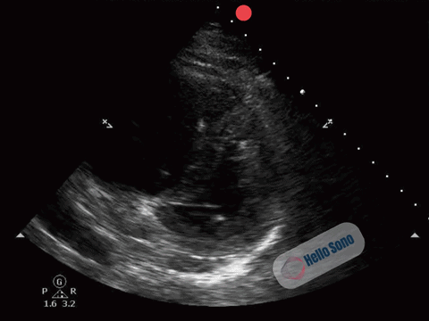
With persistent concern for pulmonary embolism (PE), the clinician proceeds with a focused cardiac POCUS exam. A representative parasternal short-axis (PSSA) view at the level of the papillary muscles is shown below.
What do you see, and what’s the diagnosis?